Periodontal disease affects ever pet that walks the planet and can develop into serious problems in up to 85% of dogs and cats by 3 years of age. Periodontal disease is initiated by bacteria that attaches in a biofilm to the pet’s teeth, called ‘plaque’. Plaque accumulation causes gum disease (gingivitis) and can be the initial step in the formation of ulcers and abscesses in your pet’s mouth. When plaque is not removed daily from the tooth surface it can harden into calculus (tartar) when bathed by saliva. When pets commence homecare programs early in life, often professional dental cleaning is not required.
Owners need to:
- Brush the pet’s teeth with a special animal toothpaste and a soft toothbrush (we recommend the Petosan range of brushes and pastes which are available from Veterinary clinics)
- Feed the pet a specially formulated dental food (we recommend the Royal Canin dental food for dogs and cats)
- Feed the pet raw meat and bones under supervision (get advice from the dental clinic as to which bones are best suited for your pet).
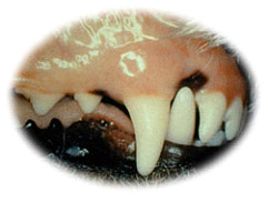
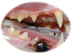
In the early stages of gingivitis, the bacterial plaque that accumulates on the tooth surface releases waste products, which damage the gums and supporting structures of the tooth, such as the jaw bone and periodontal ligament. The bacterial breakdown products also contribute to bad breath (halitosis). When a thorough professional clean is performed at this stage by a dental veterinary clinic the disease can be reversed and when homecare is provided afterwards, the health of the mouth and pet can be maintained.
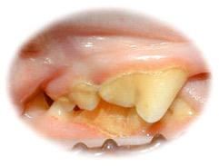
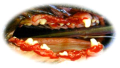
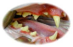
Once bacteria move under the gum further deterioration of the ligament and jaw bone occurs, the disease progresses, called ‘periodontitis’, which results in tooth mobility and eventually abscess formation and loss of the tooth. During this stage the bacteria gain access to the body’s circulation, travel to and may result in heart, liver, brain, joint and kidney disease. When bacteria have spread to the heart, liver and kidneys, supportive medication may be required. Many pet’s that have been referred to the dental clinic with advanced dental disease demonstrate increased white blood cells in their circulation and raised liver and kidney enzymes. When the disease has progressed to this stage, often the options include advanced periodontal procedures involving gum surgery, surgically raising gum tissue, placement of artificial bone materials or extraction of the teeth. When extraction is necessary, you will be warned of the consequences that this may produce, such as trauma to other parts of the mouth, future jaw fractures and the tongue falling out.

All pet’s which undergo a general anaesthesia at the dental clinic have blood and urine tests performed and a thorough physical examination completed prior to the dental procedure to ensure they are healthy. If an abnormality is found, we will discuss an alternative treatment plan. Our trained dental nurses can reschedule the surgery, work with your family Veterinarian on alternative treatments and plan the future anaesthetic and hospital visit to minimise any unforeseen situations. We have picked up many abnormalities in supposedly healthy patients with dental disease and planned and monitored the visit and anaesthesia for a successful outcome.
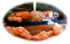
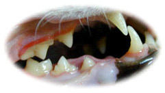
In certain breeds (Cocker Spaniels, Great Danes, Boxers, Dobermanns, Maine Coon and Abyssinian cats), bacterial plaque can lead to the gum enlarging and overgrowing the teeth. This is termed ‘gingival hyperplasia’. The enlarged gum creates a deep pseudo-pocket around the tooth which accumulates hair, food and debris resulting in further infection. A veterinary dental specialist can restore the gum to its normal healthy height by using high frequency radio waves or laser surgery. This procedure is termed ‘gingivoplasty’. The laser is used to cut the gum without bleeding and reduced pain, which restores the gum and allows the mouth’s self cleaning mechanism to work.